Articles
/Medical
CT vs MRI vs Ultrasound: Advantages and Disadvantages
This article compares the pros and cons of CT, MRI, and ultrasound scans, highlighting their strengths and limitations for medical imaging.
4 min read
Advertisement
Table of Contents
This article explores the differences between CT scans, MRI scans, and ultrasound scans. It highlights the principles behind each technology, the equipment used, their respective advantages and disadvantages.
CT stands for Computerized Tomography. This technology uses X-rays. A CT scan machine takes numerous X-ray images from different angles and a computer then constructs a 3D picture of the inside of the body.
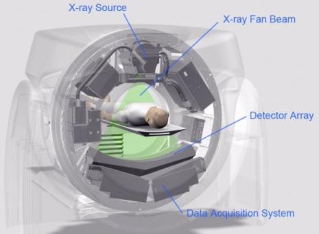
The CT scanner features a moving platform (or “cache”) where patients lie in a sleeping position. This platform slowly moves during the scanning process. Typically, two scans are performed: the first captures a general image, and the second provides a more detailed view. A complete CT scan usually takes around 5 minutes.
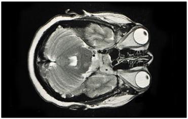
MRI stands for Magnetic Resonance Imaging. This technology relies on magnetism. It’s a more recent advancement compared to CT scans. MRI uses magnetism to create a 3D image of the body’s interior.
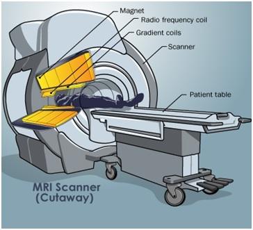
Similar to CT scanners, MRI machines have a moving platform. Patients lie down on this platform and are moved into a narrow cylinder. Magnets within the cylinder generate a strong magnetic field around the patient’s body. The cylinder is somewhat narrower compared to CT scanners. It’s crucial to remove all metallic objects from the body before an MRI scan. MRI scans can take around 30 minutes to complete.
Key components of an MRI scanner include:
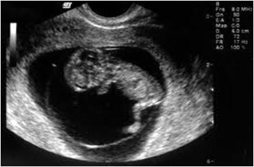
Ultrasound scans are based on ultrasound imaging and the piezoelectric effect. They are used to diagnose causes of pain, swelling, and infection in the body’s internal parts and organs. They can help in biopsies, diagnose heart conditions and assess damage after a heart attack, and are commonly used to examine babies in pregnant women.
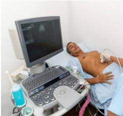
The process involves producing sound waves to create pictures of the body’s interior. Patients lie down, and a radiologist manually moves a small probe over the body to capture real-time images.
During the scan, ultrasound waves are directed into the body and reflected by tissues. These reflected waves vary in time and intensity, and a piezoelectric crystal in the probe converts the mechanical vibrations into a varying electric current. This current helps to produce an image on the ultrasound scanner’s screen.
Since ultrasound scans don’t use radiation, they are generally considered very safe and painless.
The following table summarizes the key differences between CT scans, MRI scans, and ultrasound scans:
| Feature | CT scan | MRI scan | Ultrasound scan |
|---|---|---|---|
| Waves/method used | X-Rays | Magnetic Waves | Ultrasound waves |
| Injection | Yes | Yes | No |
| Time to conduct tests | Within 5 minutes | Approx. 30 minutes | Immediate, ~5 mins analyze |
| Cost of test | Medium | High | Medium |
| Bony structure | More detail | Less detailed | Not for deep bone, upper ok |
| Soft tissue | Less detailed | Much higher detailed | Ideal |
CT scans, MRI scans and ultrasound scans each have distinct advantages and limitations, making them suitable for different medical applications.
While CT scans and MRI scans offer higher image resolution, they come with higher costs and certain risks, such as radiation exposure in CT and longer scan times in MRI. Ultrasound, though safe and portable, has limitations in imaging deep or air filled structures. The choice between these imaging techniques depends on the medical condition, patient safety and diagnostic requirements, ensuring the best possible outcome for each case.
Advertisement
Articles
/Medical
This article compares the pros and cons of CT, MRI, and ultrasound scans, highlighting their strengths and limitations for medical imaging.